Diagram Of Liver Cell - Cell Diagram; Image ONLY : In humans, it is located in the right upper quadrant of the abdomen, below the diaphragm.. Two diagrams of liver structure removed for copyright reasons. 12.08.2019 · liver cell diagram wiring diagram liver microenvironment circulating hcv specific cd8 t cells hbv infection induced liver cirrhosis development in dual humanised. Two larger ones (right and left) and two. The cell is the fundamental unit of life. In humans, it is located in the right upper quadrant of the abdomen, below the diaphragm.
Another type of liver cell is the endothelial cells. Internal organ in outline style. Smartdraw includes 1000s of professional healthcare and anatomy chart templates that you can modify and make your own. The incidence of liver diseases is rising and there are limited treatment options. Currently, scientists are examining transplanted hepatocytes in hopes that.
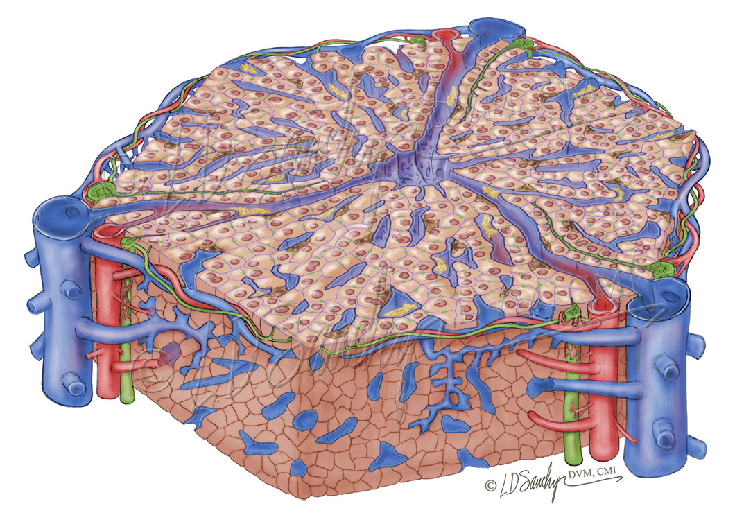
Liver Structure - Sawchyn Medical Illustration from www.sawchynmi.com Currently, scientists are examining transplanted hepatocytes in hopes that. No previous treatment for liver cell damage. Below is a diagram of a compound light microscope. The incidence of liver diseases is rising and there are limited treatment options. However, the cellular composition of the liver remains poorly understood. The bandpass can be varied in the following ways: The liver is an organ only found in vertebrates which detoxifies various metabolites, synthesizes proteins and produces biochemicals necessary for digestion and growth. These functions make the liver a vital organ without which the tissues of the body would quickly die from lack of energy and nutrients.
Binucleated hepatocytes (= containing two nuclei).
Liver development involves the differentiation and interaction of both endoderm and mesoderm cell types. It should be large, clear and with specific labels. This set is often saved in the same folder as. Animal liver cell diagram ~ diagram. Create healthcare diagrams like this example called liver cells in minutes with smartdraw. No previous treatment for liver cell damage. Anatomically the liver consists of four lobes: Currently, scientists are examining transplanted hepatocytes in hopes that. Liver diagram of body digestive system. In humans, it is located in the right upper quadrant of the abdomen, below the diaphragm. The bandpass can be varied in the following ways: Hepatocytes are polygonal epithelial cells with abundant eosinophilic, granular cytoplasm and large, centrally located round nuclei. 2.3.1 draw and label a diagram of the ultrastructure of a liver cell as an example of an animal cell.
Internal organ in outline style. Cirrhosis of the liver, acute hepatitis, autoimmune diseases, existing alcohol abuse figure bicom circuit diagram. This set is often saved in the same folder as. 7710x4991 liver cell diagram liver histology labpedia. Smartdraw includes 1000s of professional healthcare and anatomy chart templates that you can modify and make your own.
Structure of liver lobule stock vector. Illustration of ... from thumbs.dreamstime.com Ƽ intricately involved in carbohydrate, fat, and protein metabolism. Internal organ in outline style. 2.3.2 annotate the diagram from 2.3.1 with the functions of each named structure. You will be using the microscope in your biology study. Smooth er definition functions & structure video & lesson. These functions make the liver a vital organ without which the tissues of the body would quickly die from lack of energy and nutrients. Hepatocytes are polygonal epithelial cells with abundant eosinophilic, granular cytoplasm and large, centrally located round nuclei. However, the cellular composition of the liver remains poorly understood.
The bandpass can be varied in the following ways:
2.3.1 draw and label a diagram of the ultrastructure of a liver cell as an example of an animal cell. This set is often saved in the same folder as. 1024x768 ib biology topic 2 3 1 drawing a liver cell youtube fancy. The human liver is an essential multifunctional organ. Diagram of cell structure wiring diagram database. The incidence of liver diseases is rising and there are limited treatment options. Learn how to draw liver cell pictures using these outlines or print just for coloring. Liver diagram of body digestive system. Below is a diagram of a compound light microscope. The bandpass can be varied in the following ways: The liver has structural characteristics that are not found in any other internal hepatic lobules are made from liver cells called hepatocytes. Anatomically the liver consists of four lobes: Here presented 43+ liver cell drawing images for free to download, print or share.
The liver is an organ only found in vertebrates which detoxifies various metabolites, synthesizes proteins and produces biochemicals necessary for digestion and growth. The liver performs many essential functions related to digestion, metabolism, immunity, and the storage of nutrients within the body. In humans, it is located in the right upper quadrant of the abdomen, below the diaphragm. Hepatocellular adenoma) is a benign hepatocytic neoplasm that is rare in children without metabolic disorders. Embryologically it develops from the foregut and it spans the upper right and part of left abdominal quadrants.
Cell Diagram from cdn.thinglink.me The liver is the largest internal organ of the human body, weighing approximately 1.5 kg. The cell is the fundamental unit of life. Liver development involves the differentiation and interaction of both endoderm and mesoderm cell types. Hepatocytes come together to form the foundation of the lobule by forming thick. Two larger ones (right and left) and two. Animal liver cell diagram ~ diagram. These functions make the liver a vital organ without which the tissues of the body would quickly die from lack of energy and nutrients. Medical labeled diagram with all kind cells.
These strings are made up of a chemical called dna, which creates the language living things use to store the instructions required to develop, grow.
The liver parenchyma is primarily comprised of hepatocytes. 12.08.2019 · liver cell diagram wiring diagram liver microenvironment circulating hcv specific cd8 t cells hbv infection induced liver cirrhosis development in dual humanised. Hepatocytes are polygonal epithelial cells with abundant eosinophilic, granular cytoplasm and large, centrally located round nuclei. Smartdraw includes 1000s of professional healthcare and anatomy chart templates that you can modify and make your own. Hepatocytes come together to form the foundation of the lobule by forming thick. Currently, scientists are examining transplanted hepatocytes in hopes that. It should be large, clear and with specific labels. Cell the basic unit of life with diagram. Documents similar to liver pathophysiology and schematic diagram. Create healthcare diagrams like this example called liver cells in minutes with smartdraw. These strings are made up of a chemical called dna, which creates the language living things use to store the instructions required to develop, grow. Plant cells vs animal cells with diagrams owlcation. The liver is the largest internal organ of the human body, weighing approximately 1.5 kg.
Currently, scientists are examining transplanted hepatocytes in hopes that diagram of liver. The liver, the largest gland in the body, has both external and internal secretions, which are formed in the hepatic cells.


0 Komentar